
What is endoscopy nursing? The ultimate guide
Endoscopy is crucial for diagnosing and managing nearly all gastrointestinal diseases, such as gastroesophageal reflux disease, ulcers, cancer, celiac disease, and inflammation. Approximately 20 million gastrointestinal endoscopies are performed each year in the United States. Although endoscopy nurses are needed to assist with these procedures, only 254 nurses became certified gastrointestinal nurses in 2021.
Would you like to become one of these coveted gastrointestinal nurses? This ultimate guide to endoscopy nursing will cover all you need to know about working as a GI nurse.
What does endoscopy mean in medical terms?

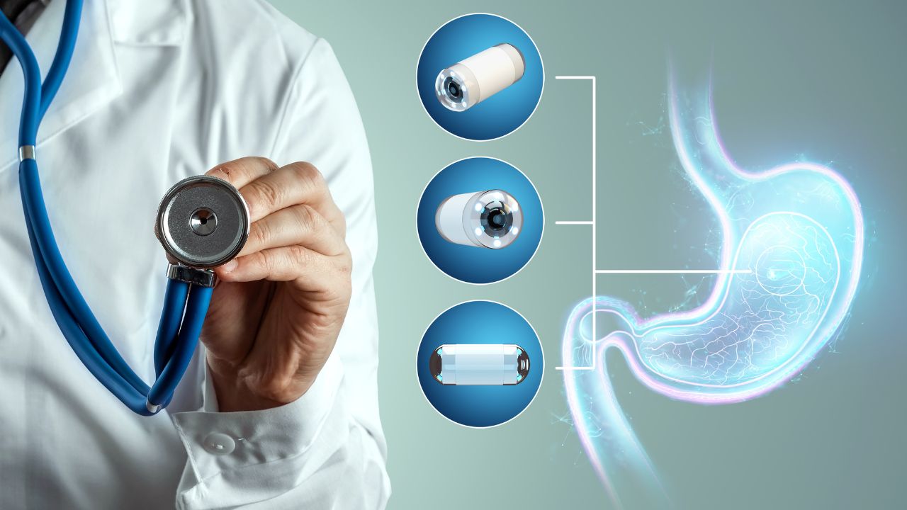
Endoscopies are minimally invasive surgical or medical procedures using an instrument called an endoscope. Endoscopes are long flexible tubes with a lens at one end and a fiber optic camera at the other, which, in conjunction, allow a magnified image to be projected onto a video screen for viewing and recording. These instruments are used to examine organs or tissue for diagnostic or therapeutic purposes. Endoscopy procedures can involve taking biopsies, dilations, retrieval of foreign objects, and removal of stones from the bile duct.
Endoscopic data from 2001 to 2005 indicates that the following are the most common endoscopy procedures:
- Colonoscopies: 61.2%
- Esophagogastroduodenoscopies (EGD): 30.6%
- Flexible sigmoidoscopies: 6.3%
- Endoscopic retrograde cholangiopancreatography (ERCP): 1%
- Endoscopic ultrasonographies (EUS): 0.8%
Although these procedures may be performed in patients of all ages, they are most common among the fifty to fifty-nine age group.
Endoscopies help to diagnose and treat symptoms and conditions affecting the esophagus, stomach, and upper intestine. The following are symptoms that endoscopies can help explain:
- Persistent heartburn
- Bleeding
- Nausea and vomiting
- Pain
- Problems swallowing
- Unexplained weight loss
GI doctors can also use endoscopies to obtain biopsies, which can be used to diagnose conditions such as cancer, celiac disease, and gastritis.
What is an endoscopy unit in a hospital?
Endoscopy units may be self-contained or share facilities with other units, such as gastroenterology, respiratory, or surgical units. Furthermore, these units may be part of hospitals, stand-alone centers, outpatient surgery centers, or self-contained rural endoscopy services.
These units usually consist of the following areas:
- A reception where patients are admitted and where family members can wait
- A preparation area, including interview facilities, patient changing rooms, and preparation rooms for pre-procedure treatments
- A procedure area
- Recovery areas
- A discharge lounge
- Staff and support areas
By definition, an endoscopy unit is dedicated to endoscopy procedures. These procedures may include all of the following:
- Gastrointestinal endoscopy, including gastroscopy, colonoscopy, and ERCP
- Endoscopic ultrasound
- Bronchoscopy
- Cystoscopy or ureteroscopy
- Duodenoscopy
- Hysteroscopy
These procedures are usually performed in controlled, sterile procedure rooms or operating rooms (ORs) using sedation or short-acting anesthetic medication. Some procedures, including ERCP, may also involve diagnostic imaging equipment. Most endoscopies are performed on a same-day basis, after which patients are either discharged or transferred to other units.
Endoscopy procedures have numerous advantages for facilities and patients:
- Procedures, recovery time, and discharge are all faster.
- Procedures are less invasive.
- Scarring is reduced.
- There is less demand for operating rooms.
What does endo stand for?
The abbreviation “endo” stands for endoscopy. Another commonly used abbreviation in this specialty is GI, which stands for gastrointestinal or gastroenterology. Gastroenterologists are often called GI doctors. Likewise, the abbreviation GI is also used to refer to gastroenterology or endoscopy nurses, who assist gastroenterologists with endoscopy procedures and care for patients with gastrointestinal symptoms or diseases in endoscopy units.
What is the role of a gastroenterology nurse?

GI nurses may assist endoscopists during endoscopic procedures and perform other tasks under the endoscopist's direct, continuous supervision and direction. These tasks include the manipulation of the endoscope and assistance with the insertion of the percutaneous endoscopic gastrostomy (PEG) tube under the principle of an “extra pair of hands.”
According to the Society of Gastroenterology Nurses and Associates, Inc. (SGNA), RNs can provide direct nursing care and technical support, whereas licensed practical nurses may only offer technical support.
Nurses assisting with endoscopic procedures must meet these general requirements:
- A licensed endoscopist must supervise nurses manipulating the endoscope and assisting with PEG tube insertion.
- Endoscopy nurses can assist endoscopists by manipulating the endoscope during endoscopic procedures.
- GI nurses should receive education regarding interventions for complications of endoscopic procedures.
- The nurse manipulating the endoscope or performing other procedures as an assistant to the endoscopist may not be the nurse monitoring the patient.
- Nurses must complete the employers’ instructional program satisfactorily and have supervised clinical practice before manipulating endoscope equipment.
- Under no circumstance should endoscopy nurses replace or assume the responsibilities of the endoscopist performing the procedure.
What does an endoscopy nurse do?
In addition to understanding the scope of practice of an endoscopy nurse, aspiring GI nurses will undoubtedly want to know what daily duties they may have to perform. Therefore, let’s take a look at some of the specific responsibilities of an endoscopy registered nurse:
- Screen patients for appropriation information
- Prepare patients for physician examination
- Wheel patients under intravenous (IV) conscious sedation to procedure rooms
- Monitor vital signs and prepare patients for exams by physicians
- Assist physicians with exams and special procedures
- Make, answer, and handle medically related phone calls as directed by physicians
- Keep exam rooms and doctors’ offices clean and maintain necessary supplies
- Assist physicians in preparing charge tickets, document fees, procedure codes, diagnosis codes, and other items as necessary
- Counsel and reassure patients, assisting them with all clinic routines
- Perform a variety of medically related tasks as directed by the physician
- Assist receptionists in scheduling appointments as needed and prioritize scheduling patients in emergency situations
- Ensure that all exam and special procedure rooms have biohazard and non-biohazard containers
- Assure that all sharps are disposed of in a Sharps Container
- Educate patients in self-administration of medication
- Chart accurately and in a timely manner
- Notify patients of test results and schedule follow-up appointments as appropriate
- Maintain patient confidentiality
- Respond to a “CODE BLUE”
- Start IVs or do IV pushes
- Mix medications, such as cytotoxic drugs
- Observe, record, and report patients’ conditions and reactions to drugs and treatments to physicians
- Dispense medication as directed
- Educate patients and family members about diagnosis procedures, medications, nutrition, and maintenance of health and wellness
How to become an endoscopy nurse?

Although the SGNS recognizes the role of LPNs in providing technical support during endoscopy procedures, most endoscopy units employ registered nurses. An aspiring ENDO nurse can become an RN in as little as two years through an associate’s degree in nursing (ADN) or four years through a bachelor’s of science in nursing (BSN) program. Although both education options are possible for endoscopy nurses, many employers prefer hiring nurses with BSNs.
In addition to RN licensure, many employers require one to two years of RN work experience in critical care, Med/Surg, or gastroenterology units.
Furthermore, most employers require Basic Life Support (BLS) and Advanced Cardiovascular Life Support (ACLS) certifications. Some employers also require IV certification.
There aren’t graduate programs for gastroenterology, but advanced certifications are available for nurses with extensive work experience in endoscopy. Nurses who wish to pursue higher education can complete an adult, family, or acute care nurse practitioner program and work in gastroenterology after graduation. Becoming a nurse practitioner takes approximately two to three additional years.
Special training to be an endoscopy nurse
Aside from completing a nursing program and obtaining work experience, nurses can pursue specialized certifications. The Accreditation Board for Specialty Nursing Certification (ABSNC) accredits the American Board of Certification for Gastroenterology Nurses (ABCGN)’s Certified Gastroenterology Registered Nurse (CGRN) credential.
According to the ABCGN, certification comes with many benefits:
- It expands a nurse’s role in providing patient services.
- It demonstrates a nurse’s commitment to their profession.
- It improves patient care.
- It helps nurses achieve greater satisfaction in their chosen profession.
- It accelerates career growth.
The CGRN credential is available for registered nurses with work experience in endoscopy. Applicants must meet the following certification criteria:
- Candidates must accrue at least two years of full-time RN work experience in GI or the part-time equivalent of 4,000 hours in the past five years.
- Candidates must submit the contact information of two responsible practitioners in the specialty who can verify their work experience and professional qualifications.
How much does an ENDO nurse make?
According to SGNA, endoscopy nurses may work in a variety of settings, including hospitals, private offices, ambulatory care centers, and clinics. This variety is significant because average salaries differ from one healthcare setting to another.
On average, LPN salary is $46,950 annually at physicians’ offices, $48,050 at general medical and surgical hospitals, and $58,170 at outpatient care centers—we’re talking about an over $10,000 difference!
For RNs, variations in salary can be even greater. On average, RN salary is $73,860 at doctors’ offices, $85,020 at hospitals, and a whopping $93,070 at outpatient care centers!
Finally, on average, advanced practice registered nurses who have become nurse practitioners (NPs) earn $114,870 at physicians’ offices, $122,960 at hospitals, and $129,190 at outpatient care centers.
What is the work environment of an endoscopy nurse like?

An important characteristic of endoscopy nursing is the schedule: Endoscopies are typically not performed at two in the morning, on Sundays, or over Thanksgiving. In other words, endoscopy nurses can usually enjoy regular waking hours as well as weekends and holidays.
“Overall, I like it. The schedule is awesome, I love having my weekends and holidays off. The only caveat I’d say is there’s not really much of a challenge and I don’t have much of an opportunity to utilize any critical thinking skills, cause usually each day is pretty typical and most cases go fine without any major issues…It’s the perfect job for someone who is feeling burnt out and wants a more laid back job in a clinical setting.” Reddit – u/taygurl
An endoscopy nurse on Reddit shared what endoscopy nursing is like depending on a nurse’s role: pre-op, intra-op, or PACU.
“First area is pre-op this is where we ask questions and start IV's…Second area is Intra-op, this is working in the room. One nurse will chart and give sedation…the second nurse will assist the physician during the procedure…Third area is PACU, this is where basically where you wake patients up, you educate them and get them out the door 30mins-1hr, depending on the patient/case. Sometimes rarely they can have breathing issues afterwards and go to the icu if in-patient. You only get a max of 2 patients. If they are ICU they skip our PACU and go straight back up to the unit.”
Is endoscopy nursing hard?
All nursing specialties have their challenges, but these can vary significantly from one specialty to the next. Whereas Med/Surg nurses are overworked and hospice nurses witness many deaths, endoscopy nurses are sometimes simply bored.
“A bad shift for me would be 40 cases and them having a bunch of advance cases like ERCP instead of simple colonoscopies or upper GI. Getting a food impaction the last 30mins of your shift or having a doctor get frustrated or yell at me. Way less stressful than working the floor imo.” Reddit – u/matt09rn
“It sounds like the complex part isn't so much the meds as much as the equipment and learning the different doctors' preferences-- everyone does something a little differently, it seems…I like admitting, it is still fast paced and busy, but the work is very repetitive. I don't mind that, but I could see how others could get bored of it.” Reddit – u/slowlymysunlight
Why choose endoscopy nursing?

Among other reasons, endo nurses love their schedules and the work-life balance that these schedules allow. Here are some testimonials from endoscopy nurses:
“Been an endoscopy nurse for 3 years and I love it! It’s my first love and I miss it so bad :( …I’m trained in the pre, intra and post procedure of endoscopy. My favorite is the intra because I do moderate sedation and help the endo techs with equipment/instruments that they need. I love the innovation and technology that come with endoscopic procedures. If you like work-life balance, go to endo.” Reddit – u/pammypies
“Love the job. A lot of variety in the units (different positions on different days). We do a fair bit of interventional work (ERCP, EUS, EBUS etc) which keeps it interesting. I also enjoy the oncall. Generally the most interesting, as well as high pressure situations (when you get major bleeds etc) occur on call ins.” Reddit – u/CochlearImplanted
What makes a good endoscopy nurse: Tips
Once you have graduated from a nursing program and passed the National Council Licensure Examination (NCLEX), you will be ready to look for nursing jobs. The following are some of the abilities, skills, and knowledge that endoscopy nursing jobs look for:
- Ability to work safely in a healthcare environment
- Ability to follow nursing procedures
- Ability to perform nursing technologies
- Ability to stand for extended periods
- Ability to problem-solve and take action on emergent events
- Being detail-oriented
- Being dependable
- Customer service skills
- Prioritization skills and ability to handle multi-tasks in a fast-paced environment
- Effective verbal and written communication skills
A great tip for obtaining more work experience to improve your resume is to pick up per diem nursing shifts. Remember that many endoscopy nursing jobs require previous work experience, so if you haven’t worked as a nurse long enough, per diem shifts can help you reach the minimum work experience requirement faster.
Final thoughts on endoscopy nursing
Endo is a great option for nurses who want to maintain circadian-rhythm-friendly hours or who value having weekends and holidays free to spend with family and friends. It is also an excellent option for nurses experiencing burnout and needing a change of pace and scenery.
Nevertheless, endoscopy nursing is not for everyone. After reading this ultimate guide, if you feel that GI nursing is not for you, continue reading about other nursing specialties until you find one that fits your strengths, preferences, and personality. Your ideal nursing specialty is out there!
